Research Article | | Peer-Reviewed

Frequency and Associated Factors of Hyperuricemia in a University Hospital in Dakar, Senegal

Received: 28 July 2026     Accepted: 8 August 2026     Published: 27 August 2026
Views:       Downloads:
Abstract

Hyperuricemia is associated with gout, cardiovascular disease, chronic kidney disease, and metabolic disorders, yet data remain limited in Senegal. This study aimed to determine the frequency of hyperuricemia and its distribution according to age, sex, and clinical indication at Fann University Hospital. This retrospective observational study was conducted from January 1 to December 31, 2025, and included patients undergoing serum uric acid testing. Hyperuricemia was defined as serum uric acid > 70 mg/L in men, > 60 mg/L in women, and > 40 mg/L in children. Associated factors were assessed using logistic regression. A total of 2,023 patients were included. The mean age was 58.5 years, and the male-to-female ratio was 0.71. The overall frequency of hyperuricemia was 29.7% and was significantly higher in men than in women (33.3% vs. 27.1%; OR = 1.34; 95% CI: 1.10-1.62; P = 0.003). Hyperuricemia increased significantly with age (OR = 1.19; 95% CI: 1.12-1.26; P < 0.001). The highest frequencies were observed in patients evaluated for cardiovascular disease (40.0%), renal disease (38.5%), and arterial hypertension (32.3%). Nearly one-third of patients had hyperuricemia, with higher frequencies in men, older individuals, and patients with cardiorenal conditions. These findings highlight the relevance of serum uric acid testing in the biological assessment of at-risk patients.

Published in Advances in Biochemistry (Volume 14, Issue 3)
DOI 10.11648/j.ab.20261403.11
Page(s) 66-71
Creative Commons

This is an Open Access article, distributed under the terms of the Creative Commons Attribution 4.0 International License (http://creativecommons.org/licenses/by/4.0/), which permits unrestricted use, distribution and reproduction in any medium or format, provided the original work is properly cited.

Copyright

Copyright © The Author(s), 2026. Published by Science Publishing Group

Keywords

Hyperuricemia, Serum Uric Acid, Cardiovascular Diseases, Senegal

1. Introduction
Uric acid (UA) is the end product of purine catabolism in humans, who lost uricase during evolution, the enzyme responsible for the oxidation of UA to allantoin, a more soluble compound that is readily excreted . Physiologically, uric acid exerts multiple biological effects, including antioxidant and pro-inflammatory activities, as well as regulatory effects on nitric oxide bioavailability and immune responses . Uric acid homeostasis depends on the balance between its production and excretion, primarily through the renal and intestinal pathways, under the influence of genetic, dietary, and environmental factors . Disruption of this balance may result in hyperuricemia, defined as an abnormally elevated serum uric acid concentration, generally above 420 µmol/L (70 mg/L) in men and 360 µmol/L (60 mg/L) in women . Hyperuricemia may be genetic or acquired and is increasingly recognized as being closely associated with several chronic conditions, including gout, cardiovascular disease, chronic kidney disease, diabetes, and obesity . Over recent decades, the prevalence of hyperuricemia has increased substantially worldwide, in both high-income and low- and middle-income countries, driven by population aging, urbanization, changes in dietary habits, and the increasing prevalence of obesity and metabolic syndrome . However, epidemiological data from sub-Saharan Africa remain limited despite the ongoing nutritional and epidemiological transitions observed in several countries . In Senegal, the few available studies have mainly focused on specific patient populations and do not provide sufficient information on the frequency of hyperuricemia among patients in hospital settings. This lack of local data limits our understanding of the burden of this biochemical abnormality and may hinder the development of appropriate screening, prevention, and management strategies. In this context, the present study aimed to determine the frequency of hyperuricemia among patients undergoing serum uric acid testing at the Biochemistry Laboratory of Fann University Hospital in Dakar, Senegal.
2. Materials and Methods
This retrospective, descriptive, and analytical study was conducted from January 1 to December 31, 2025, at the Biochemistry Laboratory of Fann University Hospital in Dakar, Senegal. The study included all patients who underwent serum uric acid testing during the study period. Patients with complete information on age, sex, and the clinical indication for serum uric acid testing were included. Patients with incomplete data and those undergoing uric acid testing as part of routine follow-up were excluded. The variables collected included sociodemographic characteristics (age and sex), requesting clinical department, clinical indication for testing, and serum uric acid concentration. Venous blood samples were collected from the antecubital vein into plain tubes. After centrifugation at 3500 rpm for 5 minutes, serum samples were analyzed immediately. Serum uric acid concentration was measured using an enzymatic uricase method on an Architect Plus ci4100 automated analyzer (Abbott Diagnostics, Illinois, USA), according to the manufacturer’s instructions. Internal quality control was performed for each analytical run to ensure the reliability of the results. Hyperuricemia was defined as a serum uric acid concentration > 70 mg/L in men, > 60 mg/L in women, and > 40 mg/L in children. Data were entered into Microsoft Excel and analyzed using Python and XLSTAT software. Continuous variables were summarized as mean ± standard deviation (SD) or median, according to their distribution, whereas categorical variables were expressed as frequencies and percentages with their 95% confidence intervals (95% CI). Continuous variables were compared using Student’s t-test or the Mann-Whitney U test, as appropriate. Categorical variables were compared using Pearson’s chi-square test or Fisher’s exact test, depending on the expected cell frequencies. Factors associated with hyperuricemia were assessed using binary logistic regression. Results are reported as odds ratios (ORs) with their corresponding 95% confidence intervals (95% CI). A two-sided P-value < 0.05 was considered statistically significant.
3. Results
After excluding 846 patients for whom the clinical indication for serum uric acid testing was either missing or could not be classified into a predefined clinical category, 2,023 patients were included in the analysis. The mean age of the study population was 58.5 ± 16.8 years, with a male-to-female ratio of 0.71. The mean serum uric acid concentration was 57.6 ± 22.2 mg/L, with a median of 53.9 mg/L (Table 1).
Table 1. General characteristics of the study population.

Parameters

Value

Sample size

2 023

Mean age

58.5 ± 16.8 years

Minimum age

2 years

Maximum age

103 years

Male-to-female ratio (M/F)

0.71

Mean serum uric acid (mg/L)

57.6 ± 22.2 (median: 53.9)

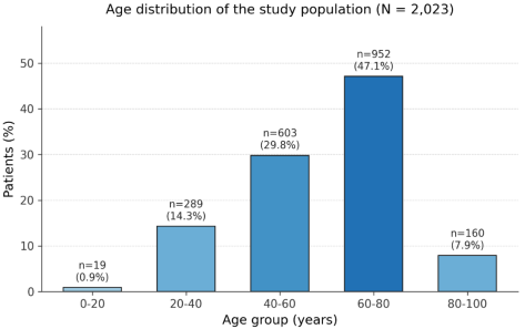
The age distribution showed a predominance of patients aged 60-80 years, accounting for 952 patients (47.1% of the study population) (Figure 1).
Figure 1. Distribution of the study population by age group.
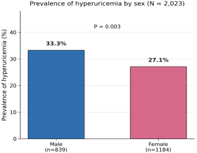
Among the 2,023 patients, 600 had hyperuricemia, corresponding to an overall frequency of 29.7%. The frequency of hyperuricemia differed significantly according to sex (P = 0.003) (Figure 2). It was significantly higher in men than in women, at 33.3% (95% CI: 30.1-36.5%) and 27.1% (95% CI: 24.7-29.7%), respectively, corresponding to an odds ratio of 1.34 (95% CI: 1.10-1.62) for men compared with women.
Figure 2. Frequency of hyperuricemia according to sex.
The frequency of hyperuricemia increased overall with age, from 20.4% among patients aged 20-40 years to 37.5% among those aged 80-100 years and older. Trend analysis, with age considered as a continuous variable, showed a significant association between increasing age and hyperuricemia (OR = 1.19; 95% CI: 1.12-1.26; P < 0.001) (Table 2).
Table 2. Distribution of hyperuricemia according to age group.

Age group, years

n

Hyperuricemia, n (%)

0-20

19

9 (47.4)

20-40

289

59 (20.4)

40-60

603

144 (23.9)

60-80

952

328 (34.5)

80-100

160

60 (37.5)

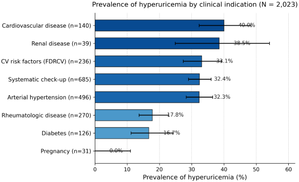
The frequency of hyperuricemia differed significantly according to the clinical indication for testing (P < 0.001) (Figure 3). The highest frequencies were observed among patients evaluated for cardiovascular disease (40.0%; 95% CI: 32.3-48.3%) or renal disease (38.5%; 95% CI: 24.9-54.1%), followed by those evaluated for cardiovascular risk factors (33.1%), undergoing routine assessment (32.4%), or evaluated for arterial hypertension (32.3%). The lowest frequencies were observed among patients with rheumatologic conditions (17.8%) and diabetes (16.7%), while no cases of hyperuricemia were observed among women undergoing pregnancy-related assessment (0/31).
Figure 3. Frequency of hyperuricemia according to clinical indication.
4. Discussion
Cardiovascular diseases are the leading cause of mortality worldwide, and hyperuricemia has attracted increasing interest because of its involvement in the development of cardiovascular, renal, and metabolic diseases. Our study provides new data on the epidemiological profile of hyperuricemia in a Senegalese hospital population by describing its overall frequency and distribution according to the main demographic and clinical characteristics. The mean age of the patients included in the study was 58.5 ± 16.8 years, with a predominance of individuals aged 60-79 years. This finding is comparable to those reported by Diack et al. in Senegal among patients with type 2 diabetes (59.6 ± 8.8 years) and by Philmon et al. in Ethiopia among hypertensive patients (57.9 ± 10.5 years) . However, unlike these studies, which focused on populations with specific chronic diseases, our study included all patients referred to the laboratory for serum uric acid testing. The predominance of older individuals probably reflects the increasing frequency of cardiovascular, renal, metabolic, and endocrine diseases with advancing age, resulting in more frequent prescription of this laboratory test . Furthermore, aging is accompanied by a progressive decline in glomerular filtration and renal urate excretion, which may contribute to increased serum uric acid concentrations. This trend may also be influenced by behavioral and therapeutic factors, including dietary habits, alcohol consumption, physical inactivity, comorbidities, and medication use, which are more common among older individuals . Women predominated in our study population, with a male-to-female ratio of 0.71. This distribution may be related to the more frequent use of healthcare services and laboratory testing by women, particularly because of their specific reproductive health needs . Serum uric acid testing is requested in various clinical settings common to both sexes, including suspected gout, renal disease, and cardiometabolic assessment. It may also be of particular interest during pregnancy in cases of gestational hypertension or pre-eclampsia, where elevated serum uric acid concentrations have been associated with adverse maternal and fetal outcomes . The mean serum uric acid concentration was 57.6 ± 22.2 mg/L, and the overall frequency of hyperuricemia was 29.7%. This frequency is close to that reported by Diack et al. in Senegal (31.1%) and by Kamdem et al. in a meta-analysis conducted in sub-Saharan Africa (31.8%) . However, it was higher than the estimates reported in an international systematic review including more than 7.4 million adults, in which the prevalence of hyperuricemia ranged from 11.2% in women to 18.6% in men between 2000 and 2023 . This difference may be explained by the hospital-based nature of our study population, which consisted of patients with clinical conditions warranting serum uric acid testing, unlike studies conducted in the general population. Consistent with previous studies , hyperuricemia was significantly more frequent in men than in women (33.3% vs. 27.1%; P = 0.003). This difference is mainly attributed to the uricosuric effect of estrogens, which reduce renal tubular urate reabsorption, particularly through the URAT1 transporter, thereby contributing to lower serum uric acid concentrations in premenopausal women . In addition to these hormonal mechanisms, metabolic and behavioral factors, such as higher consumption of purine-rich foods and alcohol and a higher prevalence of metabolic syndrome among men, may also contribute to this difference. Analysis according to clinical indication showed that the highest frequencies of hyperuricemia were observed among patients evaluated for cardiovascular disease (40.0%), followed by renal disease (38.5%) and arterial hypertension (32.1%). These findings are consistent with previous reports showing a high prevalence of hyperuricemia among patients with chronic kidney disease, cardiovascular disease, or hypertension . These associations may be explained by closely interconnected pathophysiological mechanisms. Uric acid promotes oxidative stress, reduces nitric oxide bioavailability, stimulates activation of the renin-angiotensin-aldosterone system, and contributes to chronic vascular inflammation, thereby promoting endothelial dysfunction, increased blood pressure, and vascular remodeling. Conversely, impaired renal function reduces urate excretion, creating a vicious cycle that may contribute to persistent hyperuricemia . These findings highlight the potential relevance of serum uric acid testing in patients with cardiovascular risk factors or renal impairment, both for risk assessment and laboratory monitoring. This study has several limitations. Its retrospective design precluded the collection of several variables that may influence serum uric acid concentrations, including medical history, current medications, body mass index, dietary habits, alcohol consumption, smoking status, and menopausal status. Furthermore, the single-center design may limit the generalizability of our findings to the overall Senegalese population.
5. Conclusion
This study highlights a high frequency of hyperuricemia in a Senegalese hospital population, particularly among men, older individuals, and patients with cardiovascular or renal disease. These findings suggest that identifying hyperuricemia may contribute to a more comprehensive assessment of cardiometabolic and renal risk. In the context of the ongoing epidemiological transition and the increasing burden of chronic diseases in sub-Saharan Africa, serum uric acid testing may represent a simple, accessible, and relevant tool for the screening and monitoring of at-risk patients. Further prospective, multicenter studies integrating the main clinical, biological, and environmental determinants are needed to better define the role of hyperuricemia in cardiovascular and renal prevention strategies in Senegal.
Abbreviations

UA

Uric Acid

Author Contributions
Pape Matar Kandji: Conceptualization, Data curation, Formal Analysis, Investigation, Methodology, Resources, Software, Visualization, Writing – original draft, Writing – review & editing
Moustapha Djite: Conceptualization, Data curation, Formal Analysis, Investigation, Methodology, Project administration, Resources, Software, Supervision, Validation, Visualization, Writing – original draft, Writing – review & editing
Conflicts of Interest
The authors declare no conflicts of interest.
References
[1] Roman YM. The Role of Uric Acid in Human Health: Insights from the Uricase Gene. J Pers Med. 2023; 13(9): 1409.
[2] Bartoli F, Trotta G, Crocamo C, Malerba MR, Clerici M, Carrà G. Antioxidant uric acid in treated and untreated subjects with major depressive disorder: a meta-analysis and meta-regression. Eur Arch Psychiatry Clin Neurosci. 2018; 268(2): 119-127.
[3] Kim SH, Shin J, Son HE, Kang DH. Role of urate transporters in the kidneys and intestine in uric acid homeostasis. Kidney Res Clin Pract. 2026; 45(3): 296-313.
[4] Zhang W. Hyperuricemia: Current State and Prospects. Explor Res Hypothesis Med. 2025; 10(1): 49-55.
[5] Li L, Zhang Y, Zeng C. Update on the epidemiology, genetics, and therapeutic options of hyperuricemia. Am J Transl Res. 2020; 12(7): 3167-3181.
[6] Singh G, Lingala B, Mithal A. Gout and hyperuricemia in the United States: prevalence and trends. Rheumatology (Oxford) 2019; 58: 2177-2180.
[7] Genga EK, Oyoo GO, Kalla AA. The management of gout in Africa: challenges and opportunities. Clin Rheumatol. 2021; 40: 3393-3400.
[8] Diack ND, Ndiaye N, Hedroj A, Manga AN, Dieng M, Sow D, Ndour MA, Leye YM, Diedhiou D, Sarr A, Mbaye MN, Leye A. Prevalence and associated factors of hyperuricemia in patients with type 2 diabetes in hospital settings in Dakar, Senegal. Pan Afr Med J. 2025 Jun 16; 51: 45.
[9] Philmon Daka O, Bekele Jember T, Hunie Tesfa K. Hyperuricemia and associated factors among hypertensive patients attending an academic hospital of Ethiopia: A cross-sectional study. Metabol Open. 2024; 23: 100312.
[10] Multidisciplinary Expert Task Force on Hyperuricemia and Related Diseases. Chinese Multidisciplinary Expert Consensus on the Diagnosis and Treatment of Hyperuricemia and Related Diseases. Chin Med J (Engl). 2017; 130(20): 2473-2488.
[11] Qiu L, Cheng XQ, Wu J, Liu JT, Xu T, Ding HT, et al. Prevalence of hyperuricemia and its related risk factors in healthy adults from Northern and Northeastern Chinese provinces. BMC Public Health. 2013; 13: 664.
[12] Nakanishi N, Yoshida H, Nakamura K, Suzuki K, Tatara K. Predictors for development of hyperuricemia: an 8-year longitudinal study in middle-aged Japanese men. Metabolism. 2001; 50(6): 621-626.
[13] Ni Q, Lu X, Chen C, Du H, Zhang R. Risk factors for the development of hyperuricemia: a STROBE-compliant cross-sectional and longitudinal study. Medicine (Baltimore). 2019; 98(42): e17597.
[14] WHO. Women's Health. 25 September 2018. Accessed 18 July 2026. Available online:
[15] Bresson-Just S, Houfflin-Debarge V, Herchin-Decobert A, Moranne O, Codaccioni X, Subtil D. Major elevation of uricemia associated with vomiting during the third trimester of pregnancy: five cases with favorable outcome. Gy-necol Obstet Fertil. 2004; 32(9): 713-715.
[16] Kamdem F, Doualla MS, Lekpa FK, Temfack E, Ngo Nouga Y, Sontsa Donfack O, et al. Prevalence and factors associated with hyperuricaemia in newly diagnosed and untreated hypertensives in a sub-Saharan African setting. Arch Cardiovasc Dis. 2016; 109(10): 527-532.
[17] Ngandeu-Singwe M, Nkeck JR, Hamroun A, Endomba FT, Ngoufack-Tientcheu C, Ndoadoumgue AL, Fojo BT, Ngouo AT, Tounouga DN, Nkeck JP, Youda EL, Well EA, Bigna JJ. Worldwide trends in hyperuricaemia from 2000 to 2023: a systematic review and modelling analysis. Lancet Rheumatol. 2026; 8(5): e346-e362.
[18] Lin X, Wang X, Li X, Song L, Meng Z, Yang Q, et al. Gender- and age-specific differences in the association of hyperuricemia and hypertension: a cross-sectional study. Int J Endocrinol. 2019; 2019: 7545137.
[19] Zou Y, Zhang L, Xu J, Li J, Zhang J, Long T, et al. Sex, age, and racial/ethnic disparities in hyperuricemia prevalence and risk factors among U. S. adults: an analysis of NHANES 2007-2018 data. PLoS One. 2026; 21(2): e0337714.
[20] Leslie SW, Minter DA. Hyperuricemia. 2023 Oct 14. In: StatPearls [Internet]. Treasure Island (FL): StatPearls Publishing; 2026 Jan.
[21] Rufe NB, Lamesa TA, Mamo AG, Zewude BM, Addisu B, Nigusie DJ, Biya SA. Hyperuricemia and associated factors among adult cardiovascular disease patients at Salale University Comprehensive Specialized Hospital, Fitche, Central Ethiopia. PLoS One. 2025; 20(6): e0325775.
[22] Yin Y, Zhou E, Wu J. Association between hyperuricemia and long-term mortality in patients with hypertension: results from the NHANES 2001-2018. Front Cardiovasc Med. 2024; 11: 1306026.
[23] Johnson RJ, Kang DH, Feig D, Kivlighn S, Kanellis J, Watanabe S, et al. Is there a pathogenetic role for uric acid in hypertension and cardiovascular and renal disease? Hypertension. 2003; 41(6): 1183-90.
Cite This Article
  • APA Style

    Kandji, P. M., Thioune, N. M., Djite, M., Sene, M. N., Barry, N. O. K., et al. (2026). Frequency and Associated Factors of Hyperuricemia in a University Hospital in Dakar, Senegal. Advances in Biochemistry, 14(3), 66-71. https://doi.org/10.11648/j.ab.20261403.11

    Copy | Download

    ACS Style

    Kandji, P. M.; Thioune, N. M.; Djite, M.; Sene, M. N.; Barry, N. O. K., et al. Frequency and Associated Factors of Hyperuricemia in a University Hospital in Dakar, Senegal. Adv. Biochem. 2026, 14(3), 66-71. doi: 10.11648/j.ab.20261403.11

    Copy | Download

    AMA Style

    Kandji PM, Thioune NM, Djite M, Sene MN, Barry NOK, et al. Frequency and Associated Factors of Hyperuricemia in a University Hospital in Dakar, Senegal. Adv Biochem. 2026;14(3):66-71. doi: 10.11648/j.ab.20261403.11

    Copy | Download

  • @article{10.11648/j.ab.20261403.11,
      author = {Pape Matar Kandji and Ndeye Mareme Thioune and Moustapha Djite and Moustapha Ndep Sene and Nene Oumou Kesso Barry and Mariam El Idrissi and Coumba Kamby and Youssoupha Sene and Abdou Fall and Serigne Mbacke Mbow and El Malick Ndour and Fatou Gueye-Tall and Papa Madieye Gueye},
      title = {Frequency and Associated Factors of Hyperuricemia in a University Hospital in Dakar, Senegal},
      journal = {Advances in Biochemistry},
      volume = {14},
      number = {3},
      pages = {66-71},
      doi = {10.11648/j.ab.20261403.11},
      url = {https://doi.org/10.11648/j.ab.20261403.11},
      eprint = {https://article.sciencepublishinggroup.com/pdf/10.11648.j.ab.20261403.11},
      abstract = {Hyperuricemia is associated with gout, cardiovascular disease, chronic kidney disease, and metabolic disorders, yet data remain limited in Senegal. This study aimed to determine the frequency of hyperuricemia and its distribution according to age, sex, and clinical indication at Fann University Hospital. This retrospective observational study was conducted from January 1 to December 31, 2025, and included patients undergoing serum uric acid testing. Hyperuricemia was defined as serum uric acid > 70 mg/L in men, > 60 mg/L in women, and > 40 mg/L in children. Associated factors were assessed using logistic regression. A total of 2,023 patients were included. The mean age was 58.5 years, and the male-to-female ratio was 0.71. The overall frequency of hyperuricemia was 29.7% and was significantly higher in men than in women (33.3% vs. 27.1%; OR = 1.34; 95% CI: 1.10-1.62; P = 0.003). Hyperuricemia increased significantly with age (OR = 1.19; 95% CI: 1.12-1.26; P < 0.001). The highest frequencies were observed in patients evaluated for cardiovascular disease (40.0%), renal disease (38.5%), and arterial hypertension (32.3%). Nearly one-third of patients had hyperuricemia, with higher frequencies in men, older individuals, and patients with cardiorenal conditions. These findings highlight the relevance of serum uric acid testing in the biological assessment of at-risk patients.},
     year = {2026}
    }
    

    Copy | Download

  • TY  - JOUR
    T1  - Frequency and Associated Factors of Hyperuricemia in a University Hospital in Dakar, Senegal
    AU  - Pape Matar Kandji
    AU  - Ndeye Mareme Thioune
    AU  - Moustapha Djite
    AU  - Moustapha Ndep Sene
    AU  - Nene Oumou Kesso Barry
    AU  - Mariam El Idrissi
    AU  - Coumba Kamby
    AU  - Youssoupha Sene
    AU  - Abdou Fall
    AU  - Serigne Mbacke Mbow
    AU  - El Malick Ndour
    AU  - Fatou Gueye-Tall
    AU  - Papa Madieye Gueye
    Y1  - 2026/08/27
    PY  - 2026
    N1  - https://doi.org/10.11648/j.ab.20261403.11
    DO  - 10.11648/j.ab.20261403.11
    T2  - Advances in Biochemistry
    JF  - Advances in Biochemistry
    JO  - Advances in Biochemistry
    SP  - 66
    EP  - 71
    PB  - Science Publishing Group
    SN  - 2329-0862
    UR  - https://doi.org/10.11648/j.ab.20261403.11
    AB  - Hyperuricemia is associated with gout, cardiovascular disease, chronic kidney disease, and metabolic disorders, yet data remain limited in Senegal. This study aimed to determine the frequency of hyperuricemia and its distribution according to age, sex, and clinical indication at Fann University Hospital. This retrospective observational study was conducted from January 1 to December 31, 2025, and included patients undergoing serum uric acid testing. Hyperuricemia was defined as serum uric acid > 70 mg/L in men, > 60 mg/L in women, and > 40 mg/L in children. Associated factors were assessed using logistic regression. A total of 2,023 patients were included. The mean age was 58.5 years, and the male-to-female ratio was 0.71. The overall frequency of hyperuricemia was 29.7% and was significantly higher in men than in women (33.3% vs. 27.1%; OR = 1.34; 95% CI: 1.10-1.62; P = 0.003). Hyperuricemia increased significantly with age (OR = 1.19; 95% CI: 1.12-1.26; P < 0.001). The highest frequencies were observed in patients evaluated for cardiovascular disease (40.0%), renal disease (38.5%), and arterial hypertension (32.3%). Nearly one-third of patients had hyperuricemia, with higher frequencies in men, older individuals, and patients with cardiorenal conditions. These findings highlight the relevance of serum uric acid testing in the biological assessment of at-risk patients.
    VL  - 14
    IS  - 3
    ER  - 

    Copy | Download

Author Information
  • Pharmaceutical Biochemistry Laboratory, Cheikh Anta Diop University, Dakar, Senegal; Biochemistry-Hematology Laboratory, Fann University Hospital, Dakar, Senegal

  • Biochemistry-Hematology Laboratory, Fann University Hospital, Dakar, Senegal

  • Pharmaceutical Biochemistry Laboratory, Cheikh Anta Diop University, Dakar, Senegal

  • Biochemistry-Hematology Laboratory, Fann University Hospital, Dakar, Senegal

  • Pharmaceutical Biochemistry Laboratory, Cheikh Anta Diop University, Dakar, Senegal; Biochemistry-Hematology Laboratory, Fann University Hospital, Dakar, Senegal

  • Biochemistry-Hematology Laboratory, Fann University Hospital, Dakar, Senegal

  • Biochemistry-Hematology Laboratory, Fann University Hospital, Dakar, Senegal

  • Biochemistry-Hematology Laboratory, Fann University Hospital, Dakar, Senegal

  • Biochemistry-Hematology Laboratory, Fann University Hospital, Dakar, Senegal

  • Biochemistry-Hematology Laboratory, Fann University Hospital, Dakar, Senegal

  • Pharmaceutical Biochemistry Laboratory, Cheikh Anta Diop University, Dakar, Senegal

  • Pharmaceutical Biochemistry Laboratory, Cheikh Anta Diop University, Dakar, Senegal

  • Pharmaceutical Biochemistry Laboratory, Cheikh Anta Diop University, Dakar, Senegal; Biochemistry-Hematology Laboratory, Fann University Hospital, Dakar, Senegal